CHROMOSOME: The house of genes

INTRODUCTION
Living things are made up of cells as their building blocks, within the cell are different organelles, among which is the nucleus (plural: nuclei). Inside the nucleus, the deoxyribonucleic acid (DNA) is wound tightly and packaged into a compact unit called a chromosome. So, each chromosome is comprised of DNA being tightly coiled many times around proteins named histones which act as support to its structure. Every chromosome has in it many genes, and every gene is positioned at a particular site on the chromosome, known as the locus.
DEFINITION OF CHROMOSOME
Therefore, the chromosome can now be defined as a thread-like structure of nucleic acids and proteins located in the nucleus of most living cells, which carries genetic information in the form of genes.

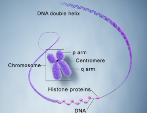
DNA and histone proteins packaged into chromosome structure (Source: U.S. National Library of Medicine).
CHROMOSOME NATURE
In eukaryotic cells (cells with nucleic region), most chromosomes are worm-shaped structurally and they are found in the nuclei. The chromosomes appear as a mass of spaghetti-like fibers with a diameter measurement of about 30 nm (30 Nanometer). In most of the larger organisms we are familiar with (as seen in humans), there are two complete homologous sets of chromosomes in each nucleus, this gives rise to a condition known as diploidy. Along the chromosome are genes in different numbers and arrangements. These genes are the functional regions along the DNA molecule that constitutes the chromosome. Those functional regions are now transcribed during the transcription process to produce RNA. The DNA region between genes which is called the intergenic segment/region shows some features or repeating patterns which is different in every chromosome. The genes on chromosomes vary enormously in size within, between, and across species. Most of these variations are caused by differences in the size and number of introns that interrupt the coding sequence of a gene called its exons). Different species have a highly characteristic number and set of chromosomes.
CHROMOSOME NUMBER
Since the number of chromosomes across species is different, it is ideal to know the chromosome number of every organism, to aid classification. To get a chromosome number, the product of two numbers should be gotten which is the haploid number (n) and the number of chromosomes in the basic genomic set. In most fungi and algae (unicellular organisms), the cells of the visible structures have only one chromosome set and therefore are called haploid. In most familiar animals and plants, the cells of the body have two sets of chromosomes; such cells are called diploid (as seen in humans) and represented as 2n.
CHROMOSOME TYPES BASED ON CENTROMERE POSITION
The region of the chromosome to which spindle fibers attach is called the centromere. In most chromosomes, it demarcates the long arm (q-arm) from the short arm (p-arm). The chromosome appears to be constricted at the centromere region and whenever the centromere is located at the end of the chromosome, the chromosome is said to be telocentric, then, the chromosome is metacentric when the centromere is in the middle. Also, a chromosome is acrocentric when the centromere is off-center (this means that the long and short arms are of unequal lengths). When there is no centromere, the chromosome is said to be acentric.
CHROMOSOME HOMOLOGY
Chromosome pair could possess the same length, centromere position, and staining pattern with genes for the same features at corresponding loci, at this point the chromosome pair is classed as being homologous (chromosome pair having similar features). Whereas, a chromosome pair with dissimilar features is non-homologous. The term chromatid describes one of the two copies of DNA that makes up a duplicated chromosome, which joins at their centromere. Those two copies are called sister chromatids when they are joined by the centromere, but once they are separated, the strands are called daughter chromatids. The end of chromosomes is called telomeres, although there is no visible structure that represents the telomere, at the end of the DNA level, rather, it is identified by identifying the end of a nucleotide sequence.
CHROMOSOME KARYOTYPE
The chromosomal constitution of any organism is described by a karyotype. The karyotype includes the total number of chromosomes, the sex chromosome constitution, and any abnormalities in number or morphology. It can also show the location of the centromere, the relative length of the short and long arms on either side of the centromere, and even the position of the constricted regions along the arms. A normal human karyotype is 46, XX for human females, and 46, XY for human males. Karyotypes are performed when cells are entering mitosis since the DNA is condensed and can be easily observed when stained.
KARYOTYPE SYMBOLS AND NOMENCLATURE
| Symbol | Meaning |
| 1-22 | Autosome number |
| Dic | Dicentric |
| Inv | Inversion |
| T | Translocation |
| Del | Deletion |
| Ins | Insertion |
| Dup | Duplication |
| :: | Break and join. |
| → | From-to |
| Mos | Mosaic |
| (+) or (-) | When placed before an autosomal number, indicates that chromosome is extra or missing. |
| X, Y | The sex chromosomes |
| P | Short arm of the chromosome |
| : | Break (no reunion, as in a terminal deletion) |
| Cen | Centromere |
| Q | Long arm of the chromosome |
| Ter | Terminal or end (pter = end of the short arm; qter = end of the long arm) |
E.g., 46, XY, del (15) (pter → q22:) represents a male with 46 chromosomes, whereby one of the 15 chromosomes has a deletion from within q22 to the qter
CHROMOSOMAL MUTATION OR ABNORMALITY
Chromosomal mutation occurs when there is a change in the number or structure of chromosomes. Such changes could result from interactions between chromosomes and mutants, and also during the processes of cell division. In mutagen-chromosome interaction, a greater dose of mutagen yields a greater number of mutations induced.
KINDS OF CHROMOSOMAL MUTATION
- Deletions: This occurs when a chromosome loses a segment as a result of breakage. The extent of lethality depends on the size of the segment lost and the coding sequence it contains. There are two kinds of deletions which are terminal deletions where the deletion removes one of the ends of a chromosome. And interstitial deletions which have the central portion of the chromosome lost.
- Inversions: This refers to rearrangements of the gene order within a single chromosome due to the inappropriate repair of two breaks. Here, the amount of chromosomal material remains unchanged, but recombination within the inverted region leads to unbalanced gametes which could lead to unbalanced miscarriage or stillbirth due to incompatibility. When inversion occurs outside the centromere of the inverted region, a paracentric inversion is reported. However, when the centromere is located within the inverted region, a pericentric inversion is reported.
- Translocations: This involves the exchange of chromosomal material between two chromosomes that are non-homologous. Approximately 4% of Down syndrome individuals are affected because of a translocation between chromosomes 14 and 21 or even between chromosomes 22 and 21.
- Duplication: Here, DNA sequences are repeated multiple times; this may result from unequal crossovers in prophase I. Note that the ratio of a gene to protein is altered if duplication produces more copies of the gene.


Good evening sir pls what is the difference between daughter chromatid and daughter chromosome
Good day dear. I guess you are referring to the same thing.. But use daughter chromosome. Because for it to be chromatic, there should be joining at the centromere
Thank you sir for the clarification