CELL DIVISION
INTRODUCTION
The growth that organisms experience is a corollary of cell division, although, it is not every cell division that leads to growth in organisms. An example is seen in eukaryotic organisms where mitotic cell division increases the number of cells of the organism. This cell number increment leads to growth and also replacement of worn-out or damaged tissue. Meiotically, these cells also divide to produce four haploid daughter cells. Such a product of four haploid cells is possible because, during meiotic cell division, the cell experiences one round of DNA replication followed by two divisions. Therefore, all cells, whether products of mitotic or meiotic cell division are proceeds of a single round of DNA replication. In meiosis, cells that are produced are dissimilar and contain as much genetic information as cells created during mitosis. This means that meiotic cell division reduces chromosomes by half. Since the cells produced are eggs and sperm cells, the zygote that results after they fertilize each other regain a full set of chromosomes. There are different stages of meiotic cell division which includes meiosis 1 and meiosis 2. Each of these stages or phases is divided further to produce other sub-stages. Meiosis 1 is acknowledged as the reduction phase because homologs are pulled to opposite poles for each pair of chromosomes but not sister chromatids whereas meiosis 2 is the division phase. At the end of the entire phase, the diploid (2n) parent cell will yield four (4) haploid gametes.
STAGES OF MEIOSIS
Meiosis can briefly be explained as a process of cell division in which the cell’s genetic information as contained in chromosomes, is mixed and divided into different sex cells with half the normal number of chromosomes that was contained in the parent cell. Each stage as stated before is divided into four further stages making it a total of eight stages. These two major stages are separated by an unusual interphase where there is no DNA synthesis or cell growth observed.
![]()
The process of meiosis I
1. Prophase 1:this stage occurs after the stage of interphase, and it is the longest stage of meiosis. It is subdivided into different stages; leptotene, zygotene, pachytene, diplotene, and diakinesis.
a) Leptotene: at this stage, the chromosome is visibly long and thin like a single thread. Also, along each of the chromosomes, small areas of thickening (chromomeres) develop which give it the expression like a necklace of beads.
b) Zygotene: at this stage, chromosomes pair and lie side by side with their pairing partner and are held together at several points along their length, creating a structure called a synaptonemal complex in the process called synapsis.
c) Pachytene: this stage commences immediately after the synaptonemal complex is formed, and it can last for days. It is characterized by the chromosome being thick and fully paired, aligning in the paired homolog to produce a distinctive pattern for each pair with the nucleoli pronounced.
d) Diplotene: this is one of the most dramatic stages because the pairing between homologs loosened a bit. So, they separate slightly but remain attached at the points where crossing over has occurred. Chiasmata (cross-shaped structure) appear between non-sister chromatids. Every of the chromosome pairs has at least one chiasma.
e) Diakinesis: here, the chromosome contracts further to a compact unit that is maximally condensed and is more maneuverable in the movements of the meiotic division.
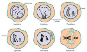
prophase 1 stages of meiosis 1
2. Metaphase 1: at this stage, the nuclear envelope has dispersed and the capture of microtubules by kinetochores occurs such that the kinetochore of sister chromatids acts as a single unit. As a result, microtubules from opposite poles become attached to the kinetochores of homologues and not to sister chromatids. Also, each joined pair of homologues line up randomly on the metaphase plate. The function of chiasmata is to help keep the pairs together. Also, it produces tension when microtubules from opposite poles attach to sister kinetochores of each homologue preparing to separate the chromatids.
3. Anaphase 1: here, the chiasmata get broken, and centromeres pull towards the pole dragging the chromosomes along with them as kinetochore microtubules shorten. As a result, one duplicated homologue goes to one pole of the cell, while the other duplicated homologue goes to the other pole. There is no separation of sister chromatids. The genes on different chromosomes assort independently into the gametes.
4. Telophase 1: this stage commences after chromosomes have segregated into two clusters with one being at each pole. Hence, the nuclear membrane re-forms around each daughter nucleus. This results in each nucleus containing two sister chromatids attached by a common centromere and the sister chromatids are no longer identical because of the crossing over that occurred in prophase 1. This increases genetic variability. At this point, cytokinesis (a division of a cell’s cytoplasm during cell division) may or may not occur while meiosis II occurs after an interval of variable length.

Stages of meiosis I
Note: some organisms like fruit fly males don’t go through recombination but they still undergo meiosis through a process called chiasmata (without chiasmata) segregation. In some species, nuclear envelopes do not re-form.
Meiosis II: between meiosis I and II is a brief interphase that does not include an S phase which is the chromosome duplication phase. Therefore, it resembles mitotic division without DNA replication. It comprises the following stages; prophase II, metaphase II, anaphase II, and telophase II.
1. Prophase II: here, spindle forms as nuclear envelopes break down.
2. Metaphase II: spindle fibers from opposite poles bind to the kinetochores of each sister chromatid and help align the chromosome along the metaphase plate in each cell.
3. Anaphase II: here, the microtubule shortens and the cohesion complex joining the centromere destroys making centromeres split as sister chromatids are pulled to opposite poles of the cells.
4. Telophase II: nuclear envelope re-forms around the four sets of daughter chromosomes thereby producing unlike nuclei content due to recombination in prophase I after which cytokinesis occurs resulting in four daughter cells that are not similar to haploid sets of chromosomes. These cells may develop directly into gametes as seen in animals or divide mitotically to produce a greater number of gametes as seen in plants, fungi, and many protists. The failure of chromosomes to move to opposite poles during any of the meiotic divisions will lead to non-disjunction and this could produce gametes lacking chromosomes and some having two copies of chromosomes. Such gametes with an improper number of chromosomes are called aneuploid gametes. This could lead to spontaneous abortion in humans.
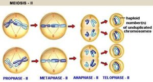
stages of meiosis II
Special features of meiosis
- Meiosis goes through homologous pairing and also crossing over to join parental homologues during meiosis I stage.
- Sister chromatids remain connected at the centromere and segregates together during anaphase I.
- The Kinetochores of sister chromatids are attached to the same pole in first division being meiosis I and to opposite poles in mitosis.
- DNA replication is suppressed between the two meiotic divisions.
- Meiosis produces cells that are not identical.
Haploid and diploid cell
Haploid cells are cells with a single chromosome set. An example is seen in gamete cells. Germ cells go through meiosis to produce 4 haploid dissimilar cells. The term haploid is applied when a cell has half the usual number of chromosomes. The number of chromosomes in a single set is represented as ‘n’. Haploid cells of human contain 23 chromosomes (n = 23). A haploid number of cells (n) differs for different organisms.
In fungi, algae, and plants, haploid spores are produced to accomplish asexual reproduction (reproduction without fusion of gametes). In male ants, they live as haploid organisms throughout their life cycle.
When cells are found with two sets of chromosomes as found in humans, they are called diploid cells. When gametes fuse at fertilization, they form a diploid zygote which in turn develops into the diploid organism. Diploid cells are the most numerous cells; nearly all the cells in the human body are diploid except gamete cells. Human has 46 chromosomes in each diploid cell hence described as 2n, meaning twice the number of chromosomes in a haploid cell (n)..
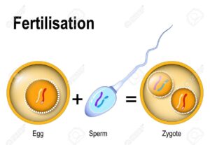
Joining of haploid gametes to form diploid zygote.
Chromosomal explanation of independent assortment
Mendel’s law of independent assortment states that different gene pairs are inherited independently during gamete formation. This law aims to explain that in a dihybrid of A/a and B/b, during gamete formation, B is probably ending up with either A or a allele in a new cell. The same applies to the b allele. But with the idea of linkage whereby genes on the same chromosome are likely to be inherited together, Mendel’s law of independent assortment has been modified and stated thus: gene pairs on separate chromosome pairs are inherited independently at meiosis.
But when the genes are on the same chromosome and close to one another, they are said to be linked and are likely to be inherited together. Also, with their recombination frequency approaching zero centiMorgan (0 cM), the greater the probability of them being assorted together, but when the recombinant frequency is approaching 50 cM, Mendel’s law may likely come to play, and at 50 cM, genes of interest assort independently.
